Home » Without Label » Left Hip Muscles Anatomy - Hip Anatomy Pictures Function Problems Treatment : The femur may also rotate around its axis about 90 degrees at the hip.
Left Hip Muscles Anatomy - Hip Anatomy Pictures Function Problems Treatment : The femur may also rotate around its axis about 90 degrees at the hip.
Left Hip Muscles Anatomy - Hip Anatomy Pictures Function Problems Treatment : The femur may also rotate around its axis about 90 degrees at the hip.. Attached to the bones of the skeletal system are about 700 named. These muscles include the gluteus maximus muscle (the largest muscle in the body) and the hamstrings group, which consists of the biceps femoris, semimembranosus, and semitendinosus muscles. Related posts of muscles of the lower back and hip diagram muscle anatomy chart. The six hip adductor muscles are all located in the adductor or medial compartment of the thigh and all mainly adduct the thigh at the hip joint. Blood vessels and nerves of the hip
Anatomy of the hip muscles. Bursae of the lower limb: Rectus femoris forms the middle portion of the quadriceps. The femur may also rotate around its axis about 90 degrees at the hip. Anterior muscles extend your legs and flex your thighs.
The Hip Anatomy On 3t Mr And 3d Pictures from www.imaios.com For detailed anatomy of pelvic bones, read anatomy of hip bone. Anterior part of the medial condyle of the tibia. Iliopsoas muscle, a hip flexor muscle that attaches to the upper thigh bone. Lateral rotation is needed for crossing the legs. Posterior view of gluteus maximus and gluteus medius in human anatomy, the muscles of the hip joint are those muscles that cause movement in the hip. Thigh magnetic resonance imaging the thigh has some of the body's largest muscles. The six hip adductor muscles are all located in the adductor or medial compartment of the thigh and all mainly adduct the thigh at the hip joint. The hip itself is a ball and socket joint, much like the shoulder.the structures necessary to create this joint are the socket, the joint capsule, muscle, ligaments, and the neck.
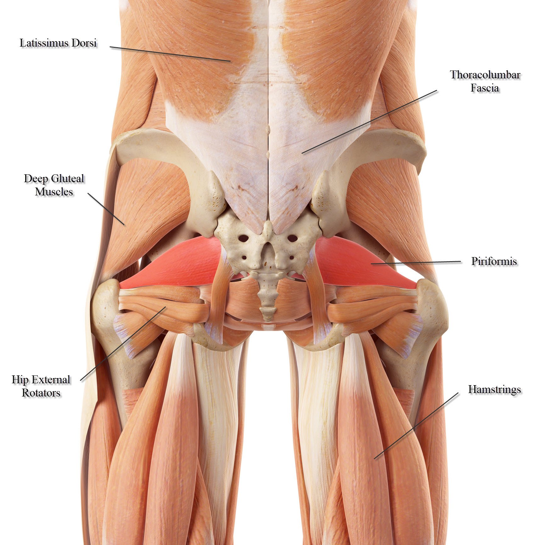
The posterior muscle group is made up of the muscles that extend (straighten) the thigh at the hip.
Hip and thigh (posterior view) if you've ever watched the videos for shakira's hips don't lie or justin timberlake's can't stop the feeling, you must've wondered how these artists can create such a wide range of hip movements.well, they have exactly the same anatomy as all of us who use those muscles to support us while we spend countless hours sitting studying the textbooks. Rectus femoris forms the middle portion of the quadriceps. Left hip muscles anatomy : Attached to the bones of the skeletal system are about 700 named. The iliofemoral, pubofemoral, and ischiofemoral ligaments represent the thickenings of the joint capsule. Injury to the iliopsoas may cause hip pain and limited mobility. The anatomy of the fascia lata and iliotibial tract; The six hip adductor muscles are all located in the adductor or medial compartment of the thigh and all mainly adduct the thigh at the hip joint. The hip joint is a ball and socket synovial type. The view on the left has the rectus femoris cut away to show the vastus intermedius which is below it. Bursae of the lower limb: The pelvic girdle (hip girdle) is formed by a single bone, the hip bone or coxal bone (coxal = hip), which serves as the attachment point for each lower limb. Some of the other muscles in the hip are:
Most modern anatomists define 17 of these muscles, although some additional muscles may sometimes be considered. This is because there are so many different muscles that give our hip joints a full range of motion. The right and left hip bones also converge anteriorly to attach to each other. Hip muscle anatomy is a complex topic. Left hip muscles anatomy :
Lower Back Muscle Anatomy And Low Back Pain from ix-cdn.b2e5.com Left hip muscles anatomy : Adductor muscles on the inside of your thigh. Lateral rotation is needed for crossing the legs. The bones together make up the hip. The muscles of the neck can be divided into groups according to. Attached to the bones of the skeletal system are about 700 named. Human anatomy hip muscles anatomy anatomy study. Hip anatomy bones and joints of the hip.
Use the mouse scroll wheel to move the images up and down alternatively use the tiny arrows (>>) on both side of the image to move the images.>>) on both side of the image to move the images.
Pick which works for you and then. Some of the other muscles in the hip are: This is because there are so many different muscles that give our hip joints a full range of motion. The right and left hip bones also converge anteriorly to attach to each other. Rectus femoris forms the middle portion of the quadriceps. Anatomy of the hip joint muscles | medicinebtg.com : The posterior muscle group is made up of the muscles that extend (straighten) the thigh at the hip. Learn about hip muscles human anatomy with free interactive flashcards. It extends from the upper arm bone to the hip bone and joins the abdominal and pectoral muscles. Injury to the iliopsoas may cause hip pain and limited mobility. Muscle anatomy chart 12 photos of the muscle anatomy chart abdominal muscle anatomy chart, human muscle anatomy diagram free, interactive muscle anatomy chart, pelvic muscle anatomy chart, shoulder muscle anatomy chart, human muscles, abdominal muscle anatomy chart, human muscle anatomy diagram free. Bursae of the lower limb: Hip muscle anatomy is a complex topic.
Area between the asis (anterior superior iliac spine) and aiis (anterior inferior iliac spine). Advanced hip flexor muscle anatomy. The femur may also rotate around its axis about 90 degrees at the hip. The hip muscles are composed of multiple flexors, extensors, adductors, abductors, and rotators that work together. Blood vessels and nerves of the hip
Psoas Major Part I Hip Flexor Or Lumbar Stabilizer from www.sportsinjurybulletin.com Pick which works for you and then. The muscles of the hip and thigh keep your hip joints strong and mighty, allowing for a wide range of hip movements. Muscle anatomy chart 12 photos of the muscle anatomy chart abdominal muscle anatomy chart, human muscle anatomy diagram free, interactive muscle anatomy chart, pelvic muscle anatomy chart, shoulder muscle anatomy chart, human muscles, abdominal muscle anatomy chart, human muscle anatomy diagram free. The strong muscles of the hip region also help to hold the hip joint together and prevent dislocation. Anatomy it band pelvis muscle pelvis with muscles hip muscles muscles of pelvis tensor fascia latae psoas major anatomy pelvis tensor fascia lata pelvis muscles. The sartorius muscle is a distinctively long and thin muscle that crosses the thigh diagonally. The thigh bone or femur and the pelvis join to form the hip joint. The six hip adductor muscles are all located in the adductor or medial compartment of the thigh and all mainly adduct the thigh at the hip joint.
The pubis, ischium, and ilium together constitute the pelvis while the thigh bone is the femur.
The hip itself is a ball and socket joint, much like the shoulder.the structures necessary to create this joint are the socket, the joint capsule, muscle, ligaments, and the neck. Anatomy of the hip joint muscles | medicinebtg.com : Hip muscle anatomy is a complex topic. This is because there are so many different muscles that give our hip joints a full range of motion. The anatomy of the fascia lata and iliotibial tract; One at the left hip, and one at the right hip. These ligaments reinforce and stabilize the hip joint(6). Some of the other muscles in the hip are: The hip's essential muscles are the sartorius, rectus femoris, gluteus minimus and medius, iliopsoas, adductors, and hamstrings. Left hip muscles anatomy : The bones together make up the hip. Injury to the iliopsoas may cause hip pain and limited mobility. The pubis, ischium, and ilium together constitute the pelvis while the thigh bone is the femur.